Figure 2.
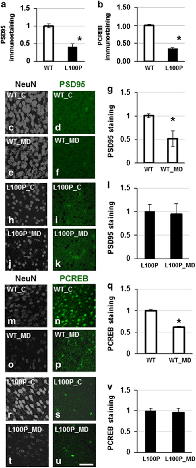
PSD95 and PCREB expression levels in WT L100P mice. (a) Measurements of PSD95 immunostaining in visual cortex sections of WT and L100P mice: the expression levels are significantly lower in the mutant. (b) The density of PCREB immunostaining significantly increases in WT but not L100P in visual cortex sections. (c–v) Immunostaining for PSD95 (c–l) shows a significant reduction of PSD95 expression in the deprived region of the visual cortex of WT (g), but not L100P (l) mice after monocular deprivation (MD). Similarly, PCREB expression (m–v) is significantly decreased in WT (q) but not L100P mice (v). Scale bar (g–j), 80 μm. *Indicates statistically significant. WT, wild type.

