Figure 5.
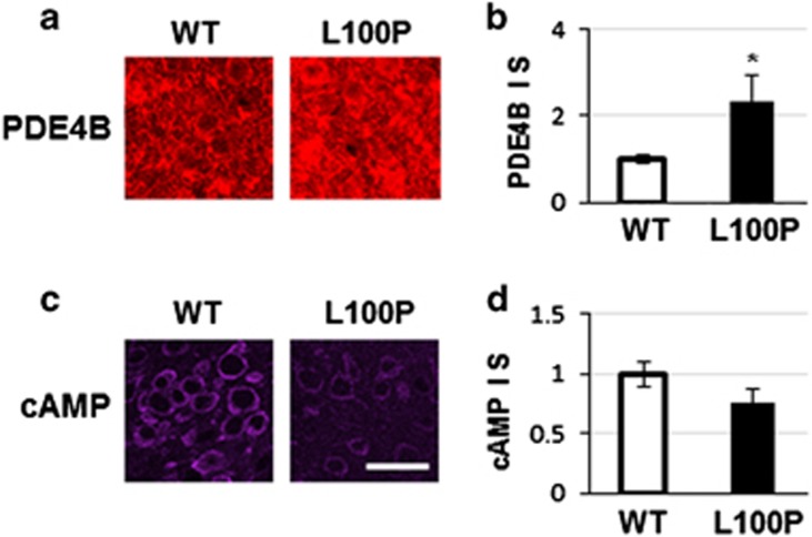
Alteration of PDE4B pathway in L100P mice. (a) PDE4B immunostaining is increased in the brain of L100P mice. Representative images from PFC. (b) Quantification of PDE4B staining across different brain regions shows significant increase of PDE4B expression in L100P mice. (c) cAMP immunostaining is decreased in the brain of L100P mice. Representative images from PFC. (d) Quantification of cAMP staining across different brain regions shows a decreased trend of cAMP expression in L100P mice. Scale bar, 40 μm. *Indicates statistically significant. PFC, prefrontal cortex; WT, wild type.

