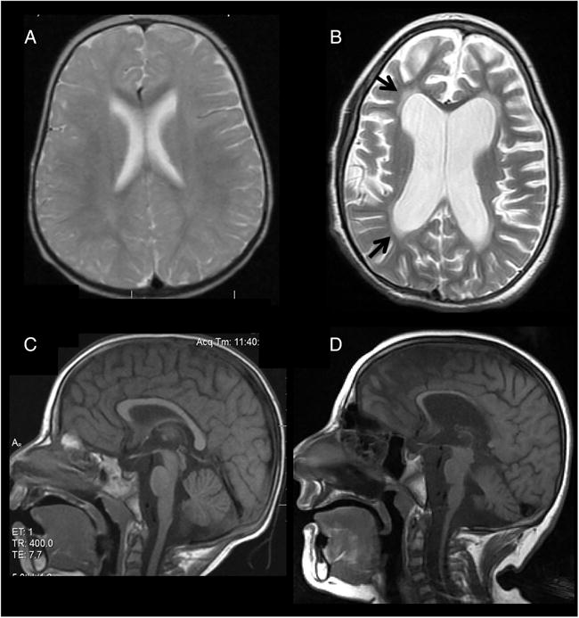
Figure 3.

Brain MRI of patient II-2 (A and B axial proton-density and T2-weighted images, respectively; C and D midsagittal T1-weighted images, respectively). Normal study is shown at age 1 year (A) and 2 years (C). At 7 years (B and D), there is diffuse supratentorial and infratentorial atrophy with thinning of the corpus callosum (D) and increased diffuse white matter signal (arrows) (B) consistent with progressive leukodystrophy.
