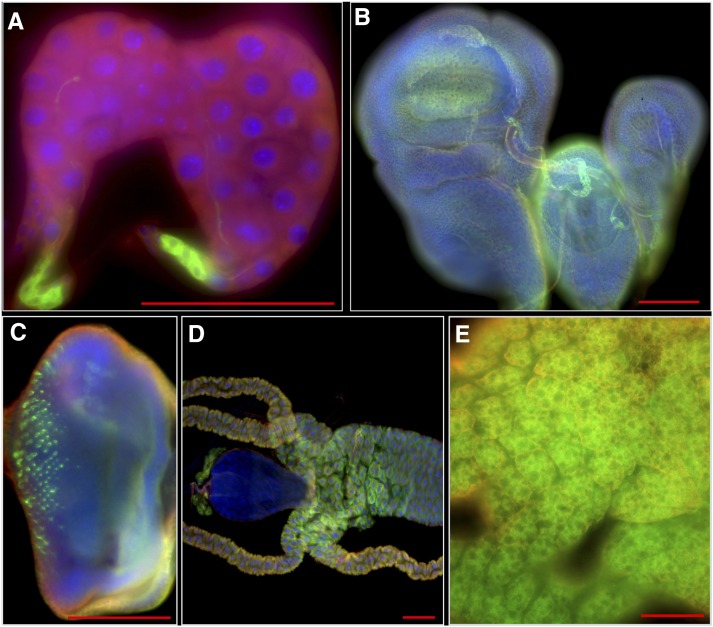
Figure 1.
Expression pattern of StanEx1 enhancer trap in tissues of wandering third instar larvae visualized by lexAop-CD8:GFP. This fly strain was used as a starter strain for the hybrid dysgenesis. For GFP channel only (green) see Figure S5. (A) CC cells in ring gland. (B) Expression in imaginal disc of wing, leg and haltere. (C) Eye disc. (D) Midgut. Note that expression in garland nephrocytes is lexAop-CD8:GFP background signal (see Materials and Methods and Figure S6). (E) Fat body. Green, Anti-GFP; Red, Anti-Tubulin; Blue, DAPI. Scale bar = 100 μm.

