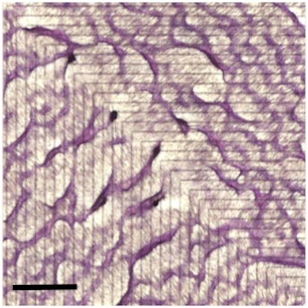
Figure 2. Macropattern formation.
Calcifying vascular cells were grown on alternating stripes of micromachined adhesive and non-adhesive substrates (fibronectin and hexamethyldisalazane/polyethylene glycol, respectively). Each stripe was 300 μm wide, and the interfaces between substrate stripes are indicated by black lines on the back (non-plated) side. In this case, a 90° bend was introduced in the configuration. After 10–14 days, the cells were fixed and stained with hematoxylin. Scale bar, 2mm.

