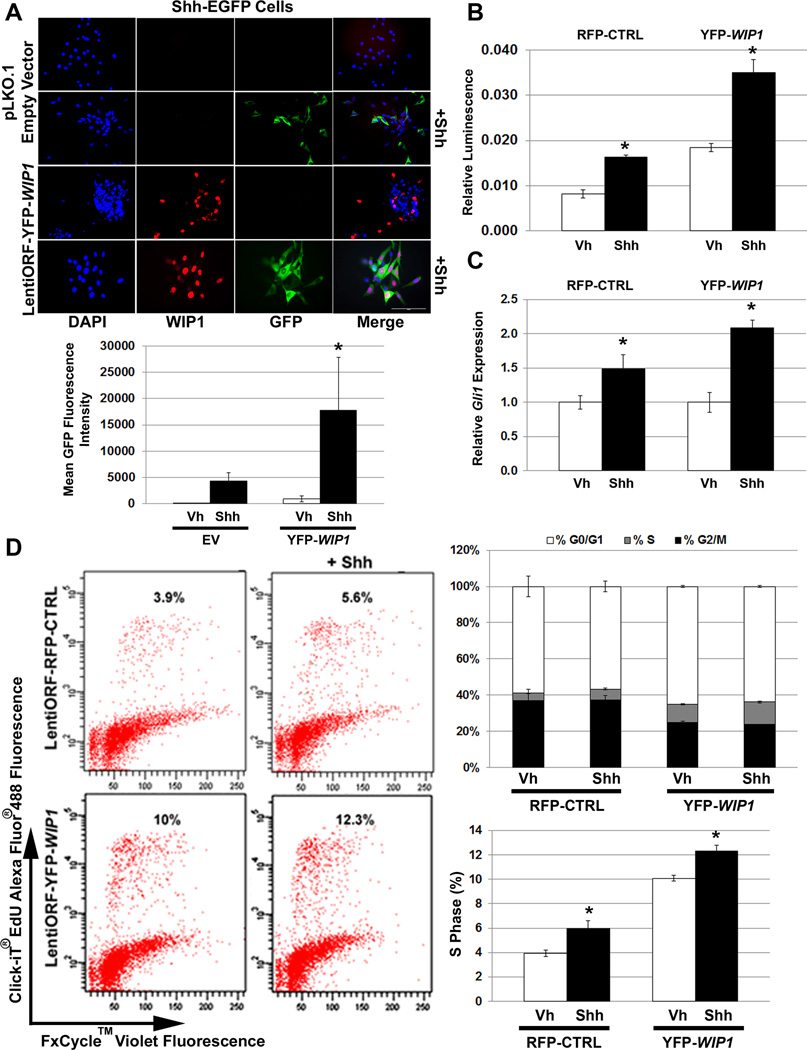
Figure 1. WIP1 promotes cell growth through hedgehog pathways.
(A) Twenty-four hours after 1×10⁵ Shh-EGFP cells were seeded on poly-L ornithine-coated plates, cells were transduced with empty vector (pLKO.1) or yellow (YFP) fluorescent protein-tagged WIP1 (YFP-WIP1) lentivirus and stimulated with vehicle (Vh) or Shh (Shh-N recombinant protein, 3µg/mL) for another 24 hours, followed by fixation and incubation with α-WIP1 primary and RFP-tagged secondary antibodies. Representative photomicrographs show fluorescence for DAPI, RFP, and GFP (top panel). Total fluorescence was measured using CellProfiler software (bottom panel), *p<0.0001. Scale bar, 100µm. (B) Shh-LIGHT2 cells were transduced with control red (RFP) fluorescent protein-tagged (RFP-CTRL) or YFP-WIP1 lentivirus and stimulated with Vh or Shh for 24 hours. Cells were subsequently lysed and assayed for expression of Firefly luciferase, relative to Renilla luciferase, *p<0.005. (C) Shh-EGFP cells, transduced with RFP-CTRL or YFP-WIP1 lentivirus and stimulated with Vh or Shh for 24 hours, were harvested and lysed for total RNA. mRNA was used to determine expression of Gli1, relative to Gapdh and normalized to expression in Vh-treated cells, by real-time, RT-PCR, *p<0.005. (D) Shh-EGFP cells were treated as in (C), then incubated with 10µM Click-iT EdU for 2 hours, followed by detection of cell cycle by flow cytometry. Error bars, standard deviation (SD) among replicates of at least three per treatment. All experiments were repeated at least three times.

