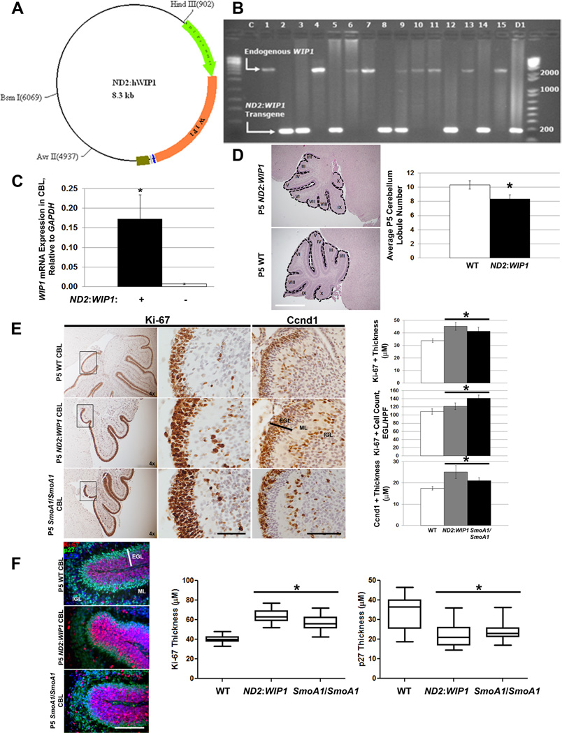
Figure 4. WIP1 promotes increased proliferation and downstream hedgehog signaling in the neonatal cerebellum.
(A) Schematic of construct used to generate ND2: WIP1 transgenic mice, showing a 1-kb portion of the Neurod2 (ND2) promoter (green arrow) driving expression of WIP1 (orange arrow; 1.8-kb). (B) DNA gel showing RT-PCR results from tail snips of ND2: WIP1 transgenic founder mice using primers spanning exons 4 and 5 of WIP1: expected 2485 base pair (bp) PCR product from endogenous Wip1; 180bp PCR product from WIP1 cDNA from expression construct in (A). C, empty template control; 1–15, founder mice; D1, PCR product using ND2: WIP1 construct as template. DNA ladders shown on either side of gel, with notations for 200–2000bp. (C) WIP1 expression by real-time RT-PCR in the cerebellum of P7 ND2: WIP1 transgenic, relative to Gapdh, compared to expression in the cerebellum of P7 wild-type (WT) C57Bl/6 mice, *p<0.005. (D) Hematoxylin and eosin stained P5 cerebella of WT and ND2: WIP1 transgenic mice (left-hand panel). Dotted line, cerebellar lobules (I–X). Quantitation of cerebellar lobules in P5 WT and ND2: WIP1 transgenic mice (right-hand panel) (n=3 cerebella per genotype), *p<0.05. (E) Immunohistochemical staining (left-hand panels) and quantitation (right-hand panels) of Ki-67 and the downstream marker of hedgehog pathway activation, Cyclin D1 (Ccnd1), in the external granule layer (EGL) of lobule X of the cerebellum of P5 wild-type, ND2: WIP1, and SmoA1/SmoA1 mice, *p<0.05. Bar, EGL width. (F) Immunofluorescent staining (left-hand panels) and quantitation (right-hand panels) of Ki-67 and p27Kip1 (p27), in the external granule layer (EGL) of the cerebellum of P5 wild-type, ND2: WIP1, and SmoA1/SmoA1 mice (n=3 cerebella per genotype), *p<0.005. Ki67, red; p27, green; DAPI, blue. Bar, EGL width. ML, molecular layer; IGL, internal granule layer; HPF, high-power field. Error bars, standard deviation (SD) among replicates of at least three per group. Scale bars, 100µm. All experiments were repeated at least three times or in at least three distinct cerebella.

