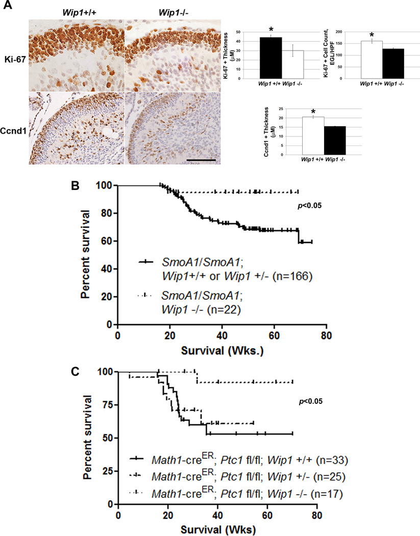
Figure 6. Wip1 knock-out suppresses MB formation in hedgehog-activated MB models.
(A) Immunohistochemical staining (left-hand panels) and quantitation (right-hand panels) of Ki-67+ and Cyclin D1 (Ccnd1)+ cells in the external granule layer of the cerebellum of P5 Wip1+/+ and Wip1−/− mice (n=3 cerebella per genotype), *p<0.005. Scale bar, 100µm. (B) Kaplan-Meier curves showing overall, MB-related survival of SmoA1/SmoA1; Wip1+/+, SmoA1/SmoA1; Wip1+/−, and SmoA1/SmoA1; Wip1−/− mice. (C) Kaplan-Meier curves showing overall, MB-related survival of Math1-creER; Ptc1 fl/fl; Wip1+/+, Math1-creER; Ptc1 fl/fl; Wip1+/−, and Math1-creER; Ptc1 fl/fl; Wip1−/− mice following treatment with 1mg tamoxifen by oral gavage per mouse at P7.

