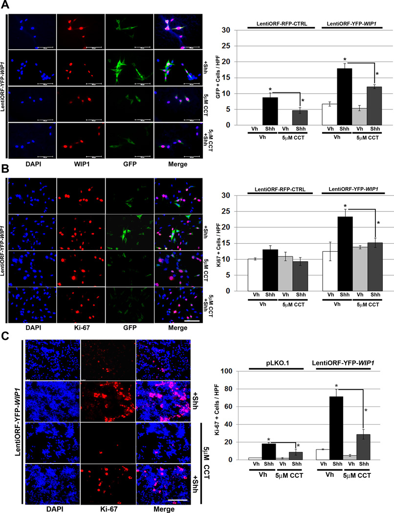
Figure 7. WIP1 inhibition suppresses hedgehog-mediated cell growth.
(A–B)Twenty-four hours after plating 1×10⁵ shh-EGFP cells, media was changed to serum-free media containing vehicle (Vh), DMSO, CCT007093 (CCT, 5µM), and/or Shh (3µg/mL). Cells were also transduced with LentiORF-RFP-CTRL or LentiORF-YFP-WIP1 lentivirus. 48 hours later, cells were fixed in 4% paraformaldehyde, permeabilized, incubated with α-WIP1 or α-Ki-67 antibody, and mounted using media containing DAPI (left-hand panels). Quantification of cells, per high power field (HPF), that express green fluorescent protein (GFP) or Ki-67 (right-hand panels). Bars, mean of cell counts from 10 representative fields for each experimental condition. (C) Twenty-four hours after plating 1×10⁶ P5 GNPs, media was changed to serum-free media containing vehicle, 5µM CCT, and/or Shh (3µg/mL). Cells were also transduced with lentivirus containing pLKO.1 or LentiORF-YFP-WIP1. 48 hours later, cells were fixed in 4% paraformaldehyde, permeabilized, incubated with α-Ki-67 antibody, and mounted using media containing DAPI (left-hand panel). Quantification of cells from (C), per high power field (HPF), that express Ki-67 (right-hand panel). Error bars, standard deviation (SD) among replicates of at least three per treatment. Scale bars, 100µm. All experiments were repeated at least three times. *p<0.005.

