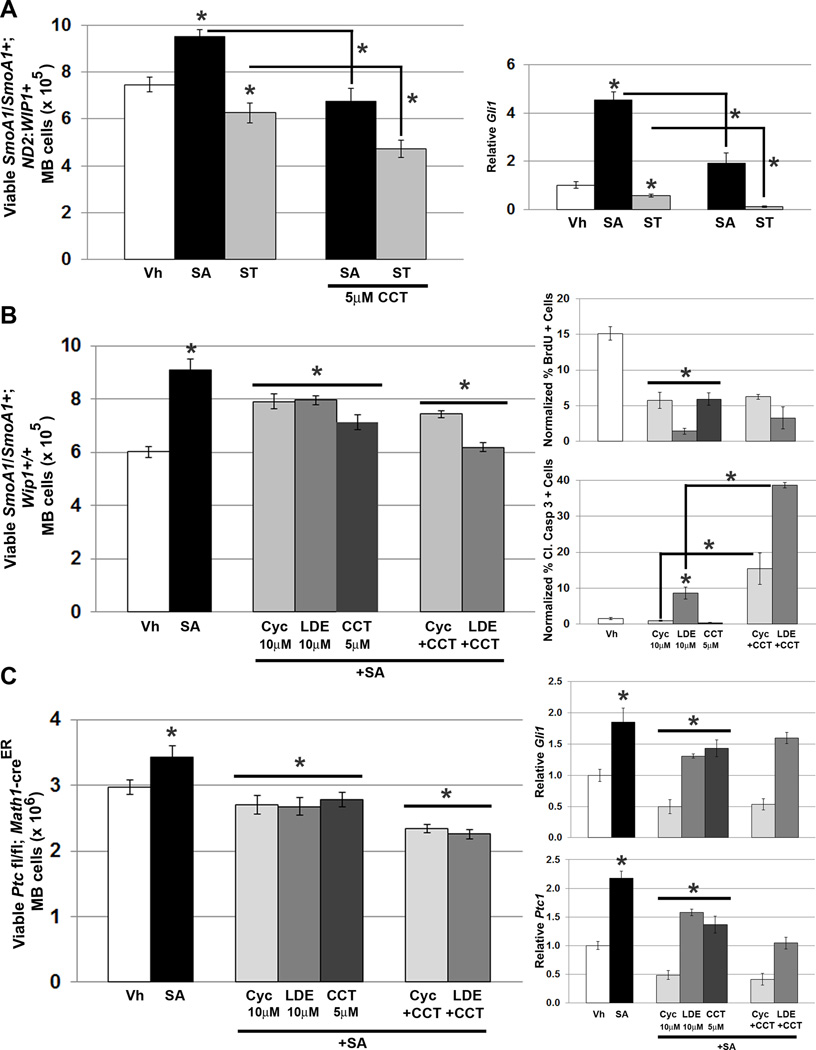
Figure 8. WIP1 inhibition augments inhibition of hedgehog signaling in hedgehog-activated MB cells.
(A). Twenty-four hours after plating 1×106 MB cells from symptomatic SmoA1/SmoA1; ND2: WIP1+ mice, media was changed to serum-free media containing vehicle (Vh), DMSO, 200nM SAG, or 100nM SANT-1, with or without CCT007093 (5µM, CCT). Viable cells were determined by trypan blue exclusion (left-hand panel), *p<0.05. Real-time, RT-PCR for Gli1, relative to Gapdh and normalized to vehicle-treated controls, using cells from (A) (right-hand panel), *p<0.05. (B) Twenty-four hours after plating 1×106 MB cells derived from symptomatic SmoA1/SmoA1; Wip1+/+ or (C) tamoxifen-induced Math1-creER; Ptc1 fl/fl mice (left-hand panels), media was changed to serum-free media containing vehicle (Vh), 200nM SAG, 10µM cyclopamine (Cyc), 10µM LDE225 (LDE), or 5µM CCT, with or without Cyc or LDE. Cells were incubated under tissue culture conditions for another 24 hours. Viable cells were determined by trypan blue exclusion (left-hand panel), *p<0.05. Real-time, RT-PCR for Gli1and Ptc1, relative to Gapdh and normalized to vehicle-treated controls, in cells from (C) (right-hand panels), *p<0.05. Medulloblastoma cells from symptomatic SmoA1/SmoA1; Wip1+/+ were also treated with Vh, 10µM Cyc, 10µM LDE, or 5µM CCT, with or without Cyc or LDE, followed by incubation with 3µg/mL BrdU for four hours, fixation in 4% PFA, permeabilization, and incubation with α-BrdU antibody, followed by incubation with Alexa Fluor 594-conjugated secondary antibody. Shown is quantitation of BrdU (B, top-right panel) or Cleaved Caspase 3 (B, bottom-right panel) immunofluorescence, relative to DAPI using CellProfiler software, *p<0.005. Error bars, standard deviation (SD) among replicates of at least three per treatment. All experiments were repeated at least three times.

