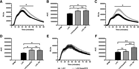
Figure 1. SaeR/S-regulated factors decrease intracellular ROS.
Human PMNs were preloaded with luminol, as described in Materials and Methods, and exposed to LAC, LACΔsaeR/S, or RPMI, and chemiluminescence was measured. (A) Time-dependent neutrophil intracellular ROS production following exposure to S. aureus LAC or LACΔsaeR/S. (B) Total relative neutrophil ROS production determined by calculating the area under the curve (AUC) from A. (C) Time-dependent neutrophil intracellular ROS production following exposure to S. aureus LAC or LACΔsaeR/S in the presence of exogenous SOD. (D) Total relative neutrophil ROS production determined by calculating the area under the curve from C. (E) Time-dependent neutrophil intracellular ROS production following exposure to S. aureus LAC or LACΔsaeR/S in the presence of exogenous catalase. (F) Total relative neutrophil ROS production determined by calculating the area under the curve from E. Data represent 5 separate experiments, using 5 different neutrophil donors; *P ≤ 0.05, **P ≤ 0.01, as determined by two-way ANOVA (A, C, and E) and one-way ANOVA (B, D, and F). RLUs, Relative luminescence units.

