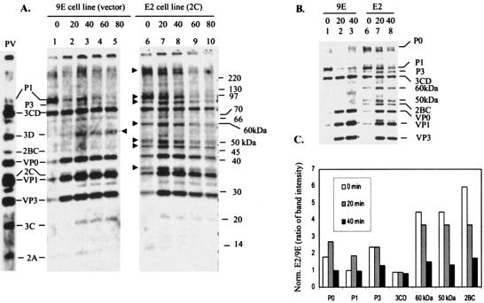
FIG. 10.
Proteolytic processing of viral precursor polypeptides in stable HeLa cell line that express 2C in an inducible fashion. (A) Pulse-chase analysis of protein processing in poliovirus-infected HeLa cells expressing the 2C polypeptide. HeLa cells lines 9E (with vector alone, lanes 1 to 5) and E2 (2C expressing cells, lanes 6 to 10) were infected at a multiplicity of infection of 10 with type 1 poliovirus as described in Materials and Methods. Cells were labeled with [35S]methionine at 3 h postinfection. The numbers above the lanes indicate times in minutes after the chase with unlabeled methionine. The lane marked PV shows the migration of poliovirus proteins from HeLa cells infected with poliovirus for 5 h. The migration of molecular markers is indicated on the right (in kilodaltons). The arrowheads pointing right indicate precursor and mature viral proteins that are either present exclusively or in higher amounts in the E2 cell line compared to the control 9E cell line. The arrowhead pointing left indicates the position of migration of 3D. (B) A lower exposure of lanes 1 to 3 and 6 to 8 from Fig. 10A is shown. (C) Quantification of viral precursor proteins from E2 and 9E cells. The viral precursor polypeptides P0, P1, P3, 3CD, 2BC, 60 kDa, and 50 kDa were quantified by densitometric scanning as described in Materials and Methods. The numerical values were normalized with respect to total cellular proteins from virus-infected E2 and 9E cells. The ratio of the numerical values for E2 to 9E cells for each polypeptide at 0, 20, and 40 min of chase are shown.

