
Figure 4. The MICAL and SelR/MsrB Reversible Redox Actin Regulatory System.
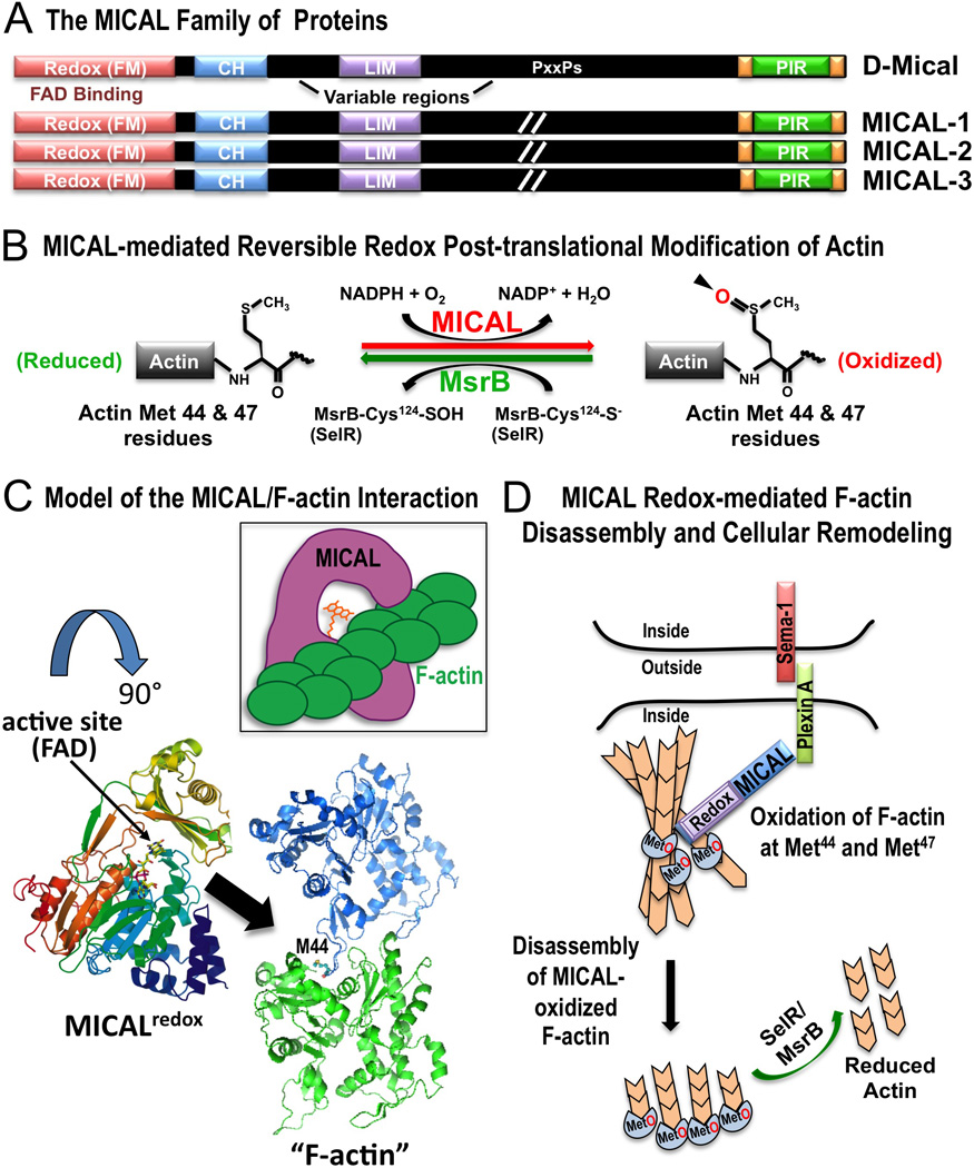
A) The MICAL family of proteins includes one Drosophila family member (D-Mical) and three vertebrate family members (MICALs 1–3) that share a conserved domain organization including the critical flavoprotein monooxygenase (Redox [FM]) domain. Each family member also contains a calponin homology (CH) domain, a LIM domain, a proline-containing (PxxPs) region, and the C-terminal plexin-interacting region (PIR). The length of the variable regions of MICAL family proteins varies and it has been designated as (//) in the diagram. (Terman et al. 2002). B) In the presence of its coenzyme NADPH, Mical adds an oxygen to both the Met-44 and Met-47 residues on F-actin. Mical oxidizes these two residues stereospecifically in the R conformation and generates actin Met-44-R-sulfoxide and Met-47-R-sulfoxide (actinMet(R)O-44 & 47) (Hung et al. 2011; Hung et al. 2013). SelR/MsrB family proteins specifically reduce MICAL-oxidized actin (Hung et al. 2013; Lee et al. 2013). C) Model of the interaction between MICAL and F-actin. The model suggests a possible mechanism by which the active site of Mical gains access to the poorly accessible Met-44 residue, which is buried within F-actin. In the model (based on published structures; PDB IDs are 2BRY, 2ZWH; (Nadella et al. 2005; Siebold et al. 2005)), rotating the Mical structure by 90 degrees (from the top of the page down) provides one mechanim to envision how the active site (small arrow) of Mical accesses (large arrow) the Met-44 (M44) residue – and as illustrated in the inset. D) Model of Mical Redox-mediated F-actin disassembly and cellular remodeling. Based on published data (Hung et al. 2011; Hung et al. 2013; Hung et al. 2010a), Semaphorin/Plexin association induces a conformational change in Plexin to allow Mical to bind to Plexin. Binding to Plexin activates Mical to associate with F-actin and oxidize the Met-44 and Met-47 residues on F-actin. Mical-mediated oxidation of F-actin induces actin filament disassembly. SelR/MsrB family proteins counteract Mical’s effects on F-actin disasembly and cellular remodeling.