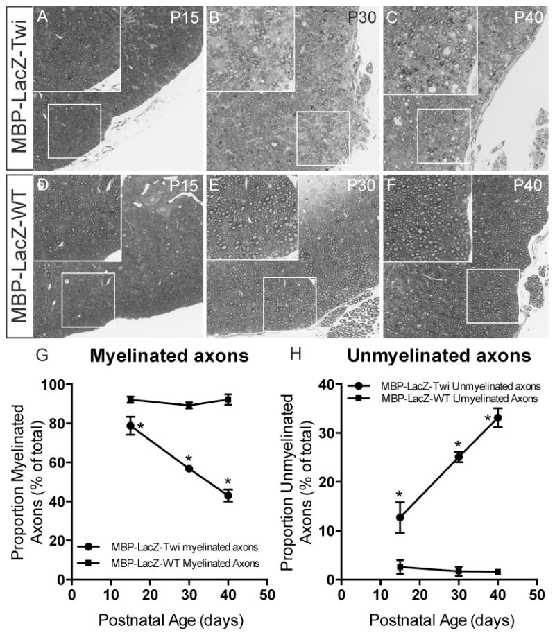
Figure 4. Demyelination of the spinal cord in MBP-LacZ-twitcher mice.

A–F) Toluidine blue staining of thin plastic sections of the lumbar spinal cord of mutant mice at P15, P30 and P40 (A–C) show progressive signs of demyelination (magnified fields in insets). G–H) The proportion of myelinated (G) and unmyelinated (H) axons as percent from the total was determined. *, p<0.05; t-test.
