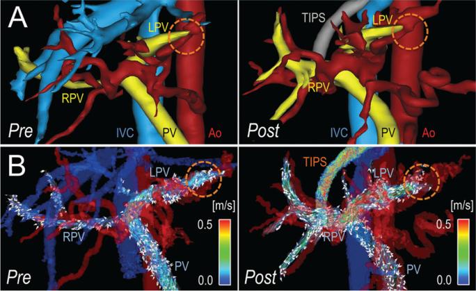
Figure 5.
Idiopathic liver cirrhosis and refractory ascites after TIPS placement in a 46-year-old man. A, Segmentation of 4D-flow angiograms obtained before (pre) and 2 weeks after (post) TIPS placement show the PV, right portal vein (RPV), left portal vein (LPV), inferior vena cava (IVC), and aorta (Ao). B, Velocity-coded 4D-flow MR images show velocity distribution in the portal system, which is indicated by color-coded streamlines that show slow flow in the PV and right portal vein (RPV) and high flow in the left portal vein (LPV). This flow pattern was caused by arterio-portal-venous shunting (dotted orange circle) that drained from a peripheral branch of the left hepatic artery and into a branch of the LPV and induced the highest measured portosystemic gradient of 28 mmHg (patient 5 in Table 1). Because of the shunt, TIPS placement further increased flow in the LPV, resulting in the fastest observed flow in the TIPS, with only a slight reduction of the portosystemic gradient to 18 mmHg. As a result, this patient had refractory ascites, even after TIPS placement.

