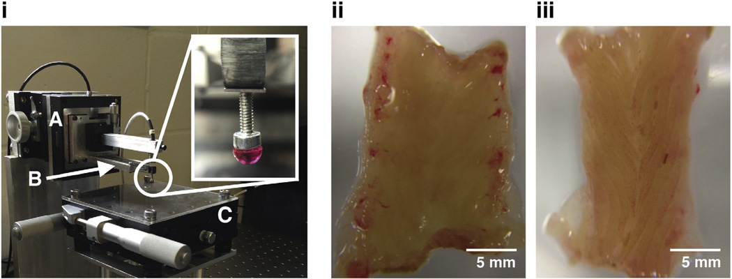
Fig. 2.
Custom-built indentation apparatus (i) used for indenting biological tissue samples. (A) Piezoelectric stage that moves cantilever base along an axis normal to the tissue surface. (B) Calibrated cantilever used for determining the normal force acting on the tip of the cantilever as the piezoelectric stages displaces the cantilever. Deflections of the cantilever are measured with a capacitive sensor. (C) X-Y stage as base for tissue sample. Micrometers enable indentations in different locations. (Inset) 3 mm-diameter rigid tip used in contact with tissue during indentation. Images of WKY (ii) and SHR (iii) colon samples isolated and cut open longitudinally for indentation. Samples were placed under the rigid tip for force-relaxation tests.

