
Fig. 1.
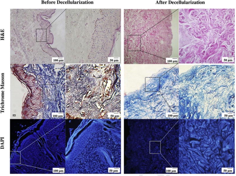
Histological assessment of human dermis before and after decellularization. H&E and Masson’s trichrome staining demonstrate that there are no cells or cellular debris and skin appendages (hair follicles, sweat glands, endocrine glands etc.) in DDM following the decellularization process. The DAPI staining shows that the DDM scaffold is free of cellular nuclei, while it retains structural integrity during processing. The Masson’s trichrome stained light micrographs illustrate that the DDM scaffolds preserved the normal collagen bundling pattern and normal collagen orientation.