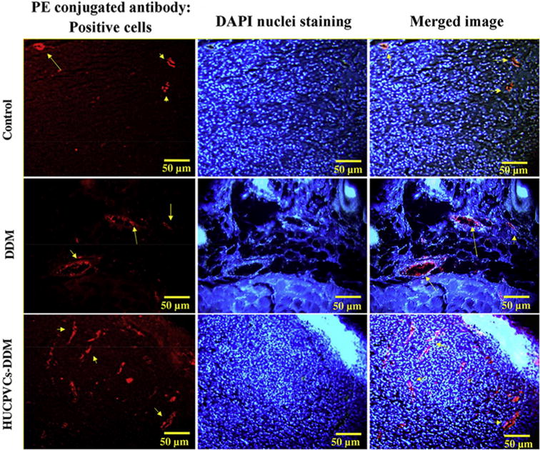
Fig. 11.

The angiogenesis activity of the scaffolds were detected by immunofluroscence staining for VEGFR-2 expression (red) in wound tissue are shown for different groups at the day 7 post implantation. The HUCPVCs-loaded DDM group showed a higher number of VEGFR-2-positive endothelial cells. (PE refers to phycoerythrin). (For interpretation of the references to colour in this figure legend, the reader is referred to the web version of this article.)
