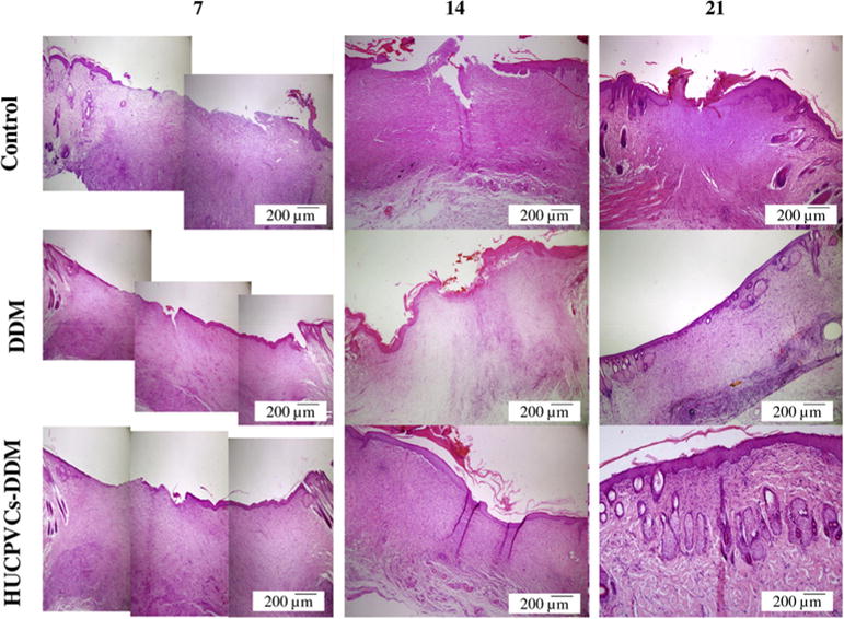
Fig. 6.

Representative photomicrographs showing the histology for the structure of epithelial and dermal layer and healing status in each group over 21 days after transplantation (H&E). After 7 days, the control, DDM and HUCPVCs loaded-DDM groups showed a number of inflammatory cells. In the HUCPVCs loaded-DDM group, some collagen fibers and fibroblasts appeared in the wound area, which related to the migration phase of wound healing process, indicating that the HUCPVCs loaded-DDM scaffold had a significant effect on the wound healing process compared with other groups. The moderate inflammation remained in the control group until 14 days. The results also show that the epidermal layer was completely formed in the HUCPVCs loaded-DDM group and covered the entire wound site after 2 weeks. However, the re-epithelization process was slow in the other groups. The HUCPVCs loaded-DDM group showed better wound healing compared with the control and DDM groups, demonstrating no sign of immunorejection over 21 days post transplantation.
