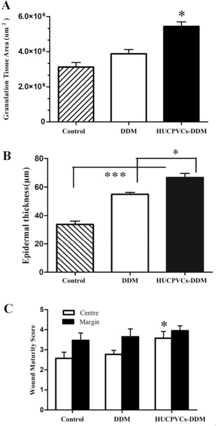
Fig. 7.

Formation of granulation tissue, epidermal thickness and wound maturity. (A) Area of granulation tissue of wounds treated with the HUCPVCs loaded-DDM scaffold were much higher than DDM and control groups (*P < 0.001). (B) The HUCPVCs-loaded DDM scaffold treated wounds demonstrated an increased thickness of the newly regenerated epidermis compared with the other groups at 7 days (*P < 0.001, ***P < 0.0001). Furthermore, the epithermal layer thickness in the DDM scaffold treated groups was significantly higher than control group (***P < 0.0001). (C) Wound maturity was measured in margin and central parts of different implanted groups, showing that the wound maturity was significantly higher in the HUCPVCs loaded-DDM treated group, especially in the central part of the wound site compared to DDM and control groups (*P < 0.001).
