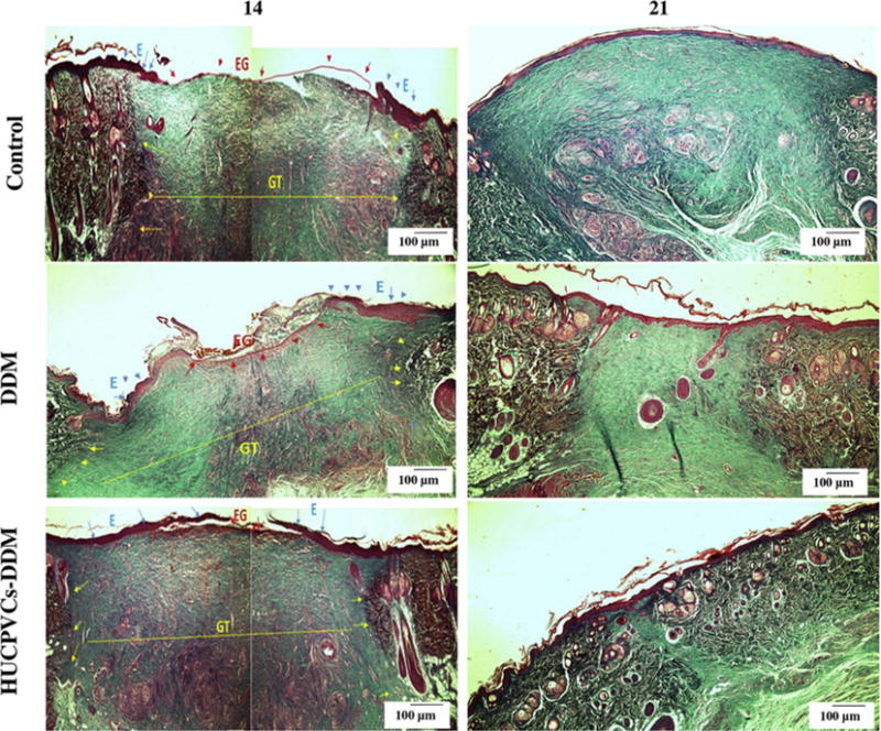
Fig. 8.

Masson’s trichrome staining of the control, DDM and HUCPVCs-DDM groups at 14 and 21 days post implantation. The collagen bundles started to appear in the wound treated with DDM and HUCPVCs-loaded DDM scaffolds after 14 days, after which a better remodeling progress was observed for the HUCPVCs-loaded DDM scaffolds. However, more collagen accumulation and deposition was observed in the control group after 21 days.
