FIG. 5.
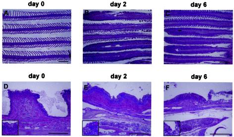
CNGV induces gill inflammation as early as 2 days p.i. Carps were infected with CNGV and harvested at the indicated days p.i. Gills were collected and subjected to histological analyses. (A to C) Gill filaments. Normally gill filaments are slender structures containing numerous lamellae (A). As early as 2 days p.i. (B), many lamellae are infiltrated by inflammatory cells. At 6 days p.i. (C) and onwards, all lamellae are heavily infiltrated. (D to F) Gill rakers. As early as 2 days p.i. (E), an increased inflammatory infiltrate is present in the subepithelial zone. In addition, at the bottom of the photomicrograph a congested vessel in the gill arch is seen. At 6 days p.i. (F), the inflammatory process is more pronounced, with sloughing of the overlying epithelium (upper right). This is accompanied by increased congestion and edema. All of the sections were stained with hematoxylin and eosin. The insets in the lower left corners are of areas in the centers of the respective photomicrograph. Bars, 200 μm.

