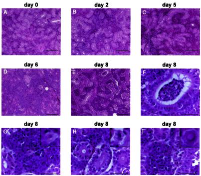
FIG. 6.
Progressive interstitial nephritis induced by CNGV. Kidneys from infected carp were collected at the indicated days p.i., and tissue sections were stained with hematoxylin and eosin. Note increased interstitial infiltration by inflammatory cells as the disease progresses. At 8 days p.i. epithelial vacuolization is also noted. (A to E) Low-power photomicrographs (bars, 100 μm). (F to I) High-power photomicrographs (bars, 40 μm). Note renal tubular inflammation (F), cytoplasmic vacuolization in a white blood cell (G), epithelial cytopathic effect (H), and a rare intranuclear inclusion body in an inflammatory cell (I). In panels G to I, the insets in the upper right corners are of cells in the centers of the respective photomicrograph.

