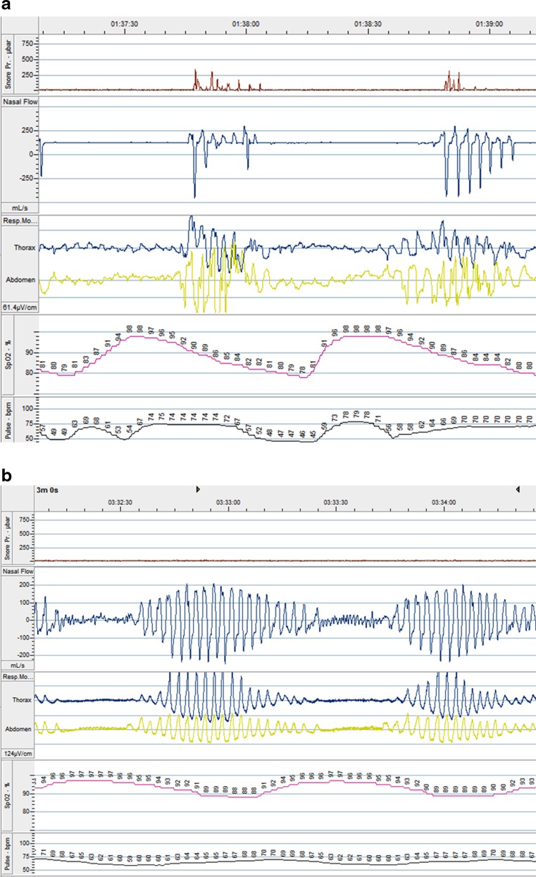
Fig. 1.
Polygraph recordings from a patient with a OSA and b CSA. Note the continuation of respiratory movement during the period of apnoea in OSA, but the absence of respiratory effort during apnoea in CSA. First panel is noise related to snoring (seen in a not b), second is nasal air flow, third is thoracic and abdominal wall movement, fourth is arterial oxygen saturation, and fifth is pulse rate (modified from reference [15])

