Figure 5.
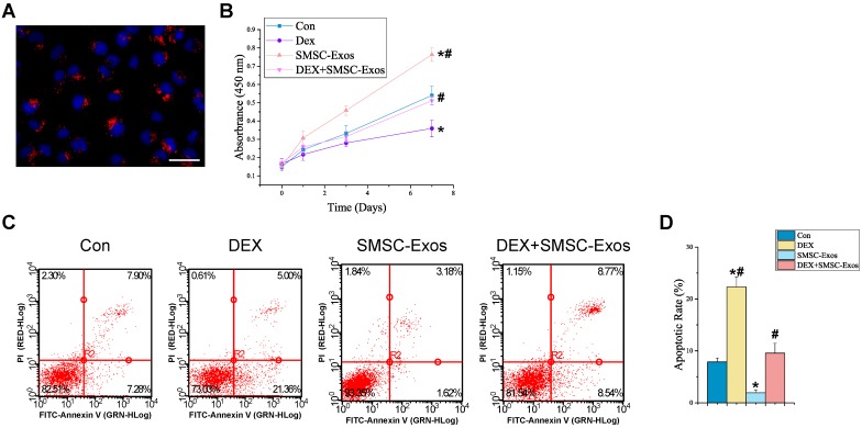
Uptake of SMSC-Exos by BMSCs and their proliferative and anti-apoptotic effects on endothelial cells. (A) Fluorescent microscopy analysis of DiL-labeled SMSC-Exos uptake by BMSCs. The red-labeled exosomes were visible in the perinuclear region of BMSCs. Scale bar: 50μm. (B) The proliferation of BMSCs was analyzed by Cell Counting Kit-8 assay. (C) The apoptosis of BMSCs was assessed by flow cytometry with annexin V-FITC/propidium iodide (PI) double staining. Cells only stained with annexin V-FITC represent the early apoptotic cells. (D) Quantitative analysis of the early apoptosis rate of BMSCs. (*P < 0.05 compared with the control group, #P < 0.05 compared with the dexamethasone (DEX)-treated group).

