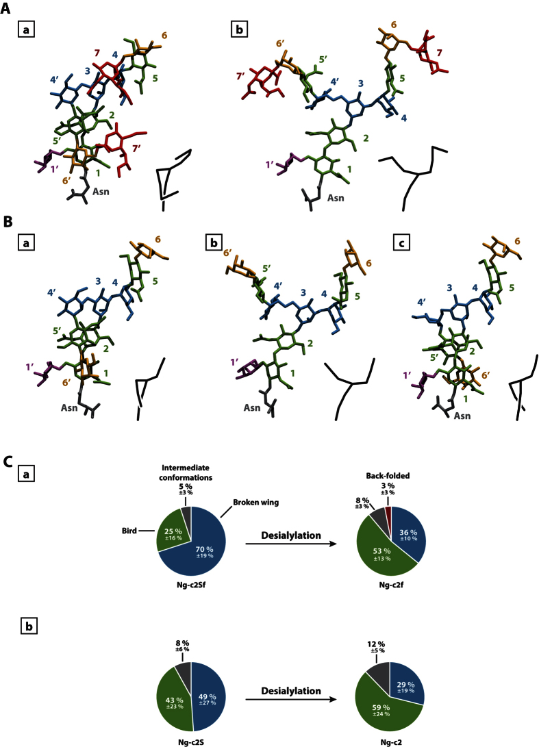
Figure 1. Identification of conformational families.
Representation of the most common –conformational states of monofucosylated disialylated bi-antennary glycan (Ng-c2Sf) (A) and monofucosylated bi-antennary glycan (Ng-c2f) (B). “Broken-wing” (a), “Bird” (b), and “back-folded”(c) conformations. The numbers refer to the numbering used in Fig. 4 and the color-codes are as follows: sialic acids in red, galactose in orange, mannose in blue, N-acetylglucosamine in green, fucose in purple, asparagine in grey. A schematic representation of the chain is provided at the bottom right of each visualization. (C) Distribution in percentage of each confor-mational state for Ng-c2Sf (a) and Ng-c2S (b) before and after the removal of sialic acids (“Broken wing” in blue, “bird” in green, “back-folded” in red, and intermediate conformations in grey).

