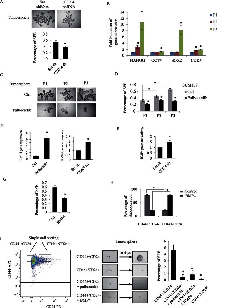
Figure 5.
(A) SUM159 cells were infected with scrambled or CDK4 shRNA overexpressing lentiviruses. Representative images of infected SUM159 cell-derived tumorspheres in the presence of 20 ng/ml EGF, 20 ng/ml bFGF, and B27 for 7 days. The number of spheres was counted and expressed as percentage of sphere-forming efficiency (SFE). (B) Tumorspheres were collected from serial passages (P1, P2 and P3). Total RNA was extracted from these three passages. Gene expression of three stem cell markers (NANOG, OCT4 and SOX2) and of CDK4 were measured by RT-PCR. (C) Representative images of tumorspheres (P1, P2 and P3) cultured in the presence or the absence of palbociclib. (D) Percentage of SFE from the indicated conditions were quantified and graphed. (E) SUM159 cells were treated with palbociclib or infected with scrambled or CDK4 shRNA. Gene expression of BMP4 was measured and graphed. (F) Scrambled or CDK4 shRNA infected SUM159 cells were transfected with BMP4 promoter or β-gal construct. Luciferase activity of BMP4 promoter construct was measured and normalized to β-galactosidase. (G) SUM159 cells were plated for tumorsphere in the presence or absence of BMP4. Percentage of SFE was measured and graphed. (H) SUM159 cells were treated with or without BMP4. The percentage of different CD44/CD24 subpopulations was analyzed and graphed by flowJo. (I) Single CD44+/CD24− and CD44+/CD24+ cell were sorted by FACSaria into 96-well low attachment plate for tumorsphere formation and cultured with or without palbociclib or BMP4 for 10 days. Percentage of SFE from the indicated conditions were quantified and graphed.

