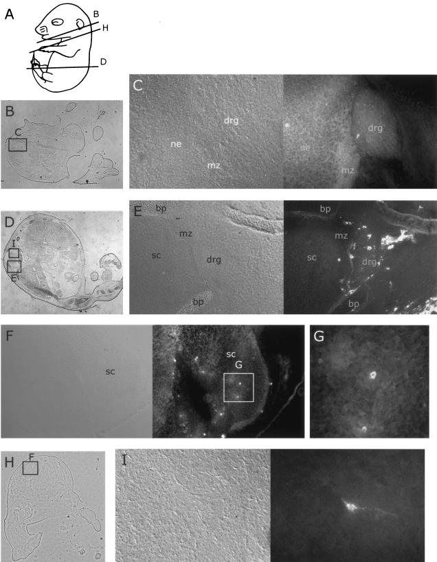
FIG. 4.
Distribution of MVM coat proteins in neural tissues of embryos inoculated at 12.5 dpc after a further 96 h of gestation. For format, see the legend to Fig. 2. (A) Approximate planes of sections in panels B, D, and H. (B) Whole section showing location of field in panel C. (C and E) Embryonic dorsal root ganglia and parts of spinal chord 96 h after inoculation with MVMi (C) or MVMp (E), showing occasional immunoreactive cells in the ganglia. (H) Whole section showing field of view in panel F. (F, G, and I) Embryonic spinal chord 96 h after inoculation with MVMi (F and G) or MVMp (I), showing occasional immunoreactive cells in the spinal chord. bp, bone primordium; drg, dorsal root ganglion; mz, marginal zone; ne, neuroepithelium; sc, spinal chord.

