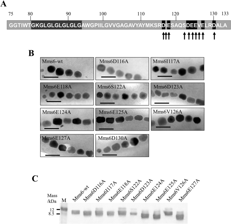
Figure 3. Characterization of Δmms6 strains harboring plasmids expressing a single amino acid substituted Mms6 protein.
(A) Location of amino acid substitutions in the Mms6 protein. The acidic or non-acidic amino acid residues substituted by alanine are indicated by black and gray arrows, respectively. (B) Transmission electron micrographs of magnetite crystals in the Δmms6 strains expressing wild-type Mms6 or amino acid mutant derivatives. Scale bar, 100 nm. (C) Western blotting analysis of His-tag fused Mms6 variants substituted with single amino acid residues. His-tag fused protein expression vectors were transformed into the Δmms6 strain. Proteins were purified from magnetosome membrane, and approximately 40 μg was loaded in each lane. M: Rainbow marker (low range).

