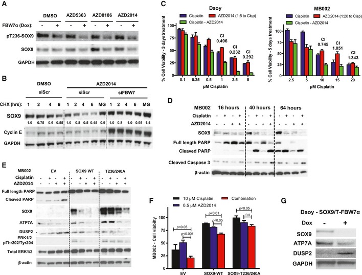
Western blotting of pT236‐SOX9 and total SOX9 following treatment of Daoy cells with a panel of inhibitors targeting PI3K/AKT/mTOR pathway. The whole‐cell lysates were collected for gel electrophoresis following 6‐h treatment with 1 μM of AZD5363, AZD8186, or AZD2014. The blots shown are representative of three independent repeats.
Western blotting of endogenous SOX9 protein turnover in the presence of PI3K/mTOR dual inhibitor, AZD2014. Daoy cells were transfected with either siScr or siFBW7 for 48 h prior to further treatment with 1 μM AZD2014. SOX9 protein turnover was examined 2 h following the inhibitor treatment by cycloheximide chase assay. Immunoblotting of cyclin E protein was used to indicate the efficacy of FBW7 knockdown. Changes in SOX9 protein level were quantified relative to GAPDH protein using ImageJ. The blots shown are representative of three independent experiments.
Quantification of resazurin‐based Daoy and MB002 cell viability following treatment with cisplatin (blue bars), AZD2014 (red bars), or their combination (green bars). Daoy cells were treated for 72 h, while MB002 cells were subjected to 5‐day course of treatment. The cell viability was calculated relative to the vehicle‐treated cells. The synthetic lethality combination index (CI) for each treatment was calculated from the mean of three independent experiments using the Compusyn software. Error bars indicate standard deviation.
Time‐course Western blotting of endogenous SOX9 and apoptotic markers including cleaved PARP and caspase‐3 following MB002 treatment with cisplatin, AZD2014, or their combination. β‐Actin was used as a loading control. The blots shown are representative of three independent experiments.
Western blotting profiling of MB002 cells expressing EV, SOX9‐WT, or SOX9‐T236/240A following 48‐h treatment with cisplatin, AZD2014, and their combination. The set of blots shown is representative of three independent experiments.
Quantification of cell viability of MB002 cells expressing EV, SOX9‐WT, or SOX9‐T236/240A following 5‐day treatment with either 10 μM cisplatin (black bars), 0.5 μM AZD2014 (blue bars), or their combination (red bars). Data are mean + standard deviation from three independent experiments, from which statistical significance was analyzed by two‐way ANOVA multiple comparisons with Bonferroni's post‐test.
Immunoblots of ATP7A and DUSP2 protein following doxycycline treatment of Daoy‐SOX9/T‐FBW7α cells. The cells were either untreated or incubated with 5 μg/ml doxycycline for 72 h to induce FBW7a prior to being harvested for gel electrophoresis. The blots shown are representative of two independent experiments.

