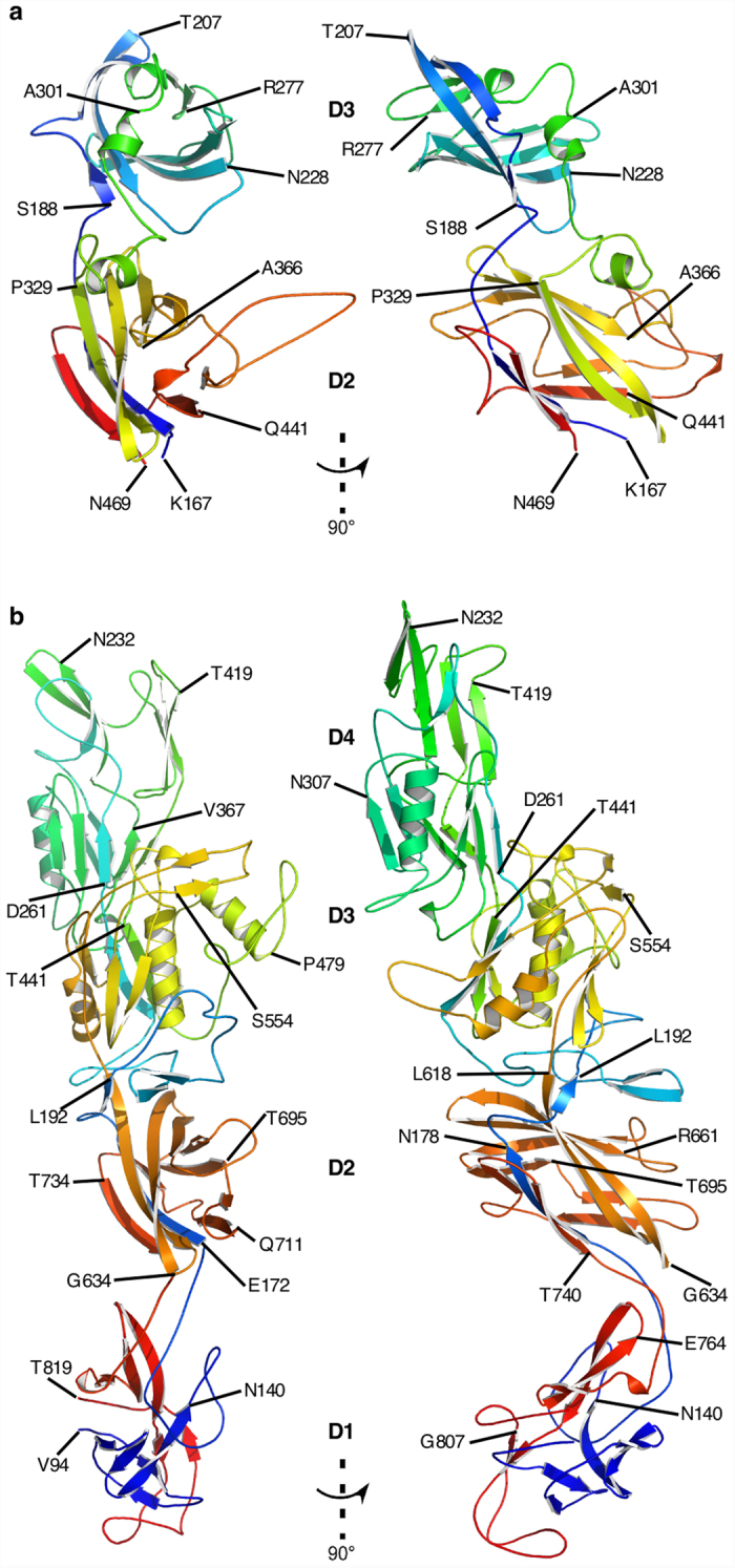
Figure 1. Structure of FlgE proteins from C. crescentus and C. jejuni.

(a) Views of the Cα backbone of FlgEcc32 with its 2 domains, D2 and D3. (b) Two different views of the Cα backbone of FlgEcj79 with its 4 domains, D1, D2, D3, and D4. The chains are colour-coded from the N- to the C-termini in rainbow colour sequence from blue to red.
