Figure 2.

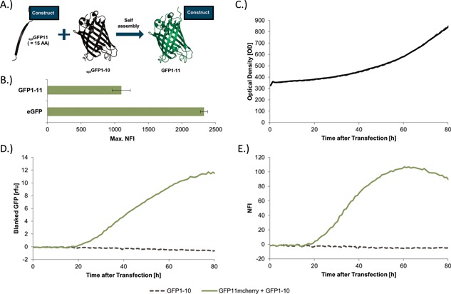
Establishment of the SplitGFP system in insect cells. (A) Schematic representation of the SplitGFP principle. One β‐strand of splGFP is fused to the protein of interest. If splGFP1‐10 is co‐expressed in the same cells, a fully active splGFP1‐11 will reassemble automatically without further interaction partners. (B) Comparison of the maximal measured fluorescence of eGFP and full‐length splGFP1‐11 in transfected Hi5 cells in the BioLector. (C) The diagram shows two Hi5 cultures, one transfected with splGFP1‐10 and the second with splGFP1‐10 and splGFP11‐mCherry. The OD for both cultures increased simultaneously. (D) In contrast, the measured GFP fluorescence only increases, when both splGFP11mCherry and splGFP1‐10 are present in the cells. Cells solely transfected with splGFP1‐10 did not show a GFP response. (E) The normalized splGFP fluorescence intensity (NFI) indicates the overall expression normalized by transfection efficacy and measured OD. The max. NFI for splGFP11mCherry was reached after 50–60 htp.
