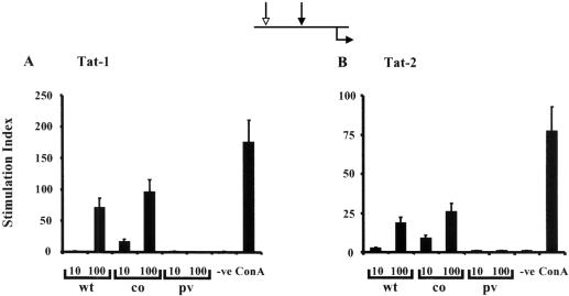
FIG. 3.
Tat-specific lymphoproliferative immune response. C57BL/6 mice (four mice per group) were genetically immunized with two different doses of Tat-1 (A) or Tat-2 (B) expression vector. The line diagram at the top of the figure depicts the immunization regimen; the white arrow represents the primary immunization, the black arrow represents the booster immunization, and the black arrow below the line represents the time of harvest. Splenocytes were harvested 1 week after the booster immunization. In a 24-well plate, 5 × 10⁶ primed splenocytes were incubated with recombinantly expressed antigen, His-Tat-1, or GST-Tat-2 at a concentration of 5 μg/ml for 5 days. Following incubation, the cells were incubated with 5 μCi of [3H]thymidine per ml for 3 h at 37°C. A stimulation index above 3 was considered a positive response. Results are presented as the means of three samples ± 1 standard deviation (error bars), and the data are representative of two independent experiments. The mice had been immunized with 10 or 100 μg of wild-type Tat (wt), codon-optimized Tat (co), or parental vector (pv). The cells were grown in the absence (-ve) and presence of ConA as a control for cell proliferation.

