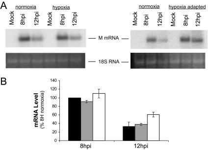
FIG. 2.
Viral mRNA accumulation during infection of hypoxic cells. (A) Total RNA was prepared from HeLa cells mock infected (M) or infected with VSV for 8 and 12 hpi. RNA was separated on a 1% gel and transferred to nitrocellulose, and viral mRNA was detected by using a 32P-labeled probe for M mRNA. Phosphorescence images of representative gels comparing infected normoxic and hypoxic cells (left) or normoxic and hypoxia-adapted cells (right) are shown at the top, with an acridine orange stain showing 18S RNA at the bottom. (B) Quantification of accumulation of M mRNA. Black bars, normoxic infection; gray bars, hypoxic infection; white bars, hypoxia-adapted infection. Values are normalized to normoxic infection at 8 hpi (8H). Values are means ± standard deviations of the results from three separate experiments.

