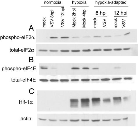
FIG. 4.
eIF-2α and eIF-4E phosphorylation during VSV infection under normoxic or hypoxic conditions. Extracts from mock- and VSV-infected cells were analyzed by Western blotting (see Materials and Methods) with antibodies against total and phospho-eIF-2α (Ser51) (A), total and phospho-eIF-4E (Ser209) (B), and the hypoxia-induced protein HIF1α and actin (C). Blots are representative of the results from two separate experiments.

