Figure 12.

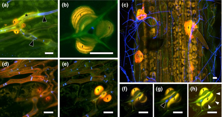
Wound response of Lolium perenne to Epichloë festucae ∆noxA mutant. (a) WGA‐AF488 fluorescence indicative of chitin (black arrow heads) in endophytic hyphae and plant autofluorescence (yellow) indicative of phenolic compounds in anticlinal epidermal cell walls (‘1’) as well as wound response between the mesophyll and epidermis (asterisk). (b) Enlargement of (a) showing several layers of cell wall deposition (probably cellulose and lignin). (c) Numerous wound responses in anticlinal epidermal cell walls and cell wall depositions (‘1’). (d–g) Ascending substacks of an endophytic hypha penetrating between epidermal cells showing plant autofluorescence indicative of a plant wound response in the anticlinal epidermal cell walls close to mesophyll (‘1’) and on top of the periclinal epidermal cell walls (‘2’). (h) Overexposed image from z‐stack of (d–g), highlighting the plasmalemma (white arrowheads) indented by plant cell wall thickening. Confocal laser scanning microscopy images of aniline blue/WGA‐AF488 costained blade sample. Bars, 10 μm.
