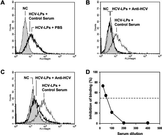
FIG. 2.
Antibody-mediated inhibition of cellular HCV-LP binding. HCV-LPs were incubated with anti-HCV-positive serum or a pool of anti-HCV-negative control sera (dilution, 1:50). HCV-LP-antibody complexes were added to HuH-7 cells for 1 h at 4°C. After removal of nonbound HCV-LP-antibody complexes by washing the cells in PBS-2% BSA, the binding of HCV-LPs was detected by flow cytometry using the mouse monoclonal anti-E2 antibody AP33 and PE-conjugated anti-mouse IgG (A and B) or human polyclonal anti-HCV and FITC-conjugated anti-human IgG antibody (C). The fluorescence intensity and relative cell numbers (Counts) are shown on the x and y axes, respectively. NC, negative control, corresponding to HuH-7 cells incubated with control insect cell preparations (GUS) and control serum. (D) For the assessment of concentration-dependent antibody-mediated inhibition of binding, HCV-LPs (genotype 1b) were incubated in subsaturating concentrations with an anti-HCV-positive serum or a pool of anti-HCV-negative control sera (at various dilutions as indicated on the x axis) for 1 h at 37°C. HCV-LP-antibody complexes were added to HuH-7 cells for 1 h at 4°C. HCV-LP binding to the HuH-7 cells was detected by flow cytometry as described above. Inhibition of cellular HCV-LP binding (y axis) was calculated as described in Materials and Methods.

