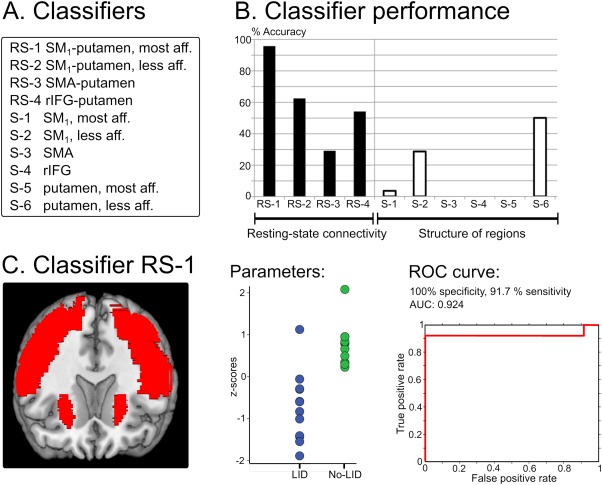
Figure 2.

Classifier analyses. A: List of classifiers. B: Classifier accuracy in distinguishing patients with and without levodopa‐induced dyskinesias (LID). C: Key parameters of the best classifier (SM1‐putamen connectivity in most affected hemisphere). AUC, area under the curve; rIFG, right inferior frontal gyrus; ROC, receiver‐operating characteristic; RS, resting state; SM1, primary sensorimotor cortex; S, structural. [Color figure can be viewed in the online issue, which is available at wileyonlinelibrary.com.]
