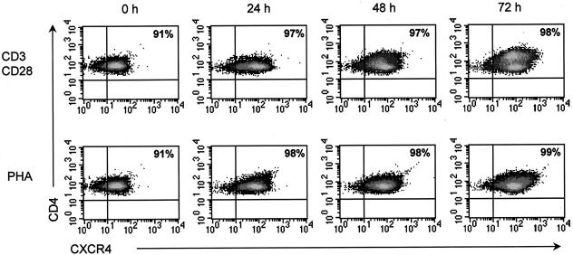
FIG. 3.
CD3/CD28-costimulated CD4+ T cells express high levels of CXCR4. CD4+ T cells were isolated from PBMC of three donors and then stimulated with CD3/CD28 antibodies or PHA for 0, 24, 48, or 72 h. At each time point, cells were stained with fluorescently conjugated monoclonal antibodies directed against CD4 and CXCR4. Samples were acquired on a FACSCalibur instrument (Becton Dickinson), and the resulting data were analyzed using CellQuest software (Becton Dickinson). Representative results from one donor are shown. The number in the upper-right hand corner of each FACS plot represents the percentage of cells that express both CD4 and CXCR4.

