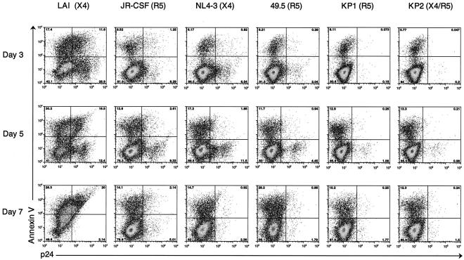
FIG. 6.
The percentage of costimulated CD4+ T cells infected by each viral strain was determined by intracellular p24 staining. CD3/CD28-costimulated CD4+ cells were infected with LAI, JR-CSF, NL4-3, and 49.5 at an MOI of 0.01 and with KP1 and KP2 at an MOI of 0.001. Apoptotic cells were detected by surface staining with fluorescently conjugated annexin V, and infected cells were detected by intracellular staining for HIV-1 p24 antigen. Samples were acquired on a FACSCalibur instrument (Becton Dickinson), and the resulting data were analyzed using FlowJo software (Tree Star, Inc.). Results from days 3, 5, and 7 postinfection are shown. The number in each corner of each FACS plot represents the percentage of cells in that quadrant.

