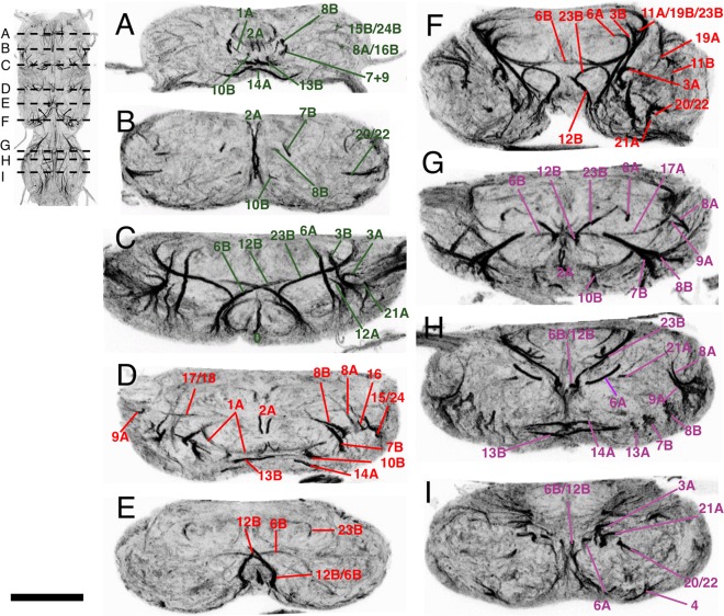
Figure 2.

Transverse sections of the adult VNS neuroglian‐labeled hemilineage scaffold in the adult VNS. Each panel is a 5‐μm‐thick optical slice through the same confocal stack at a position to give the most complete image of the scaffold organization. The sequence of images (A–I) moves from anterior to posterior with the position of each section shown in the inset figure. (A–C) Prothoracic neuromere. (D–F) Mesothoracic neuromere. (G–I) Metathoracic neuromere. Dorsal is up. Scale bar = 100 μm.
