Figure 8.

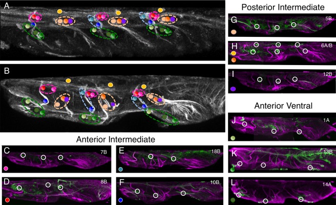
Identifying the larval commissures established by larval hemilineage in the adult VNS. (A) Sagittal section (10 μm) through the larval VNS labeled to reveal the neuroglian scaffold. (B) Sagittal section (10 μm) through the adult VNS labeled to reveal the neuroglian scaffold. The positions of the commissural processes of the postembryonic lineages are indicated with colored spots. The color of the spot indicates which lineage forms that commissure. The collective larval commissures are indicated by the dotted lines encircling the individual projections. (C–L) Sagittal sections (10 μm) through double‐labeled adult VNS showing the neuroglian scaffold (magenta) and the structure of the hemilineage revealed by GFP expression (green). (C–F) The lineages forming the larval anterior intermediate commissure. The lineages anterior to lineage 2 (10B and 18B) are in the blue circle and lineages posterior to lineage 2 (7B and 8B) are in the red circle. (G–I) The lineages forming the larval posterior intermediate commissure in the orange circle. (J–L) The lineages forming the larval anterior intermediate commissure in the green circle.
