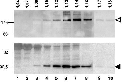
FIG. 4.
Incorporation of SARS S protein into MLV particles. Pelleted virus particles were layered on top of a 20 to 60% sucrose gradient and centrifuged to equilibrium. The gradient was fractionated from the top, and individual aliquots were trichloroacetic acid pelleted and then analyzed for the presence of HA-tagged CΔ19 (arrowhead in upper panel) and MLV Gag/p30 (arrowhead in lower panel) by Western blotting with monoclonal anti-HA or polyclonal anti-Gag antibody. The density of each fraction, in grams per milliliter, is shown. Molecular sizes of marker proteins (in kilodaltons) are indicated on the left.

