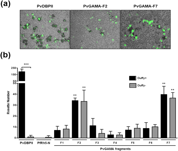
Figure 5. Binding specificity of PvGAMA fragments expressed on HEK 293T cells to Duffy-positive and -negative erythrocytes.
(a) Erythrocyte-binding rosettes formed on the surface of HEK 293T cells expressing PvDBPII or different fragments of PvGAMA were visualized under light microscopy. (b) The number of rosettes formed by the HEK 293T cells transfected with genes coding for either PvDBPII, the non-binding domain of PfRH5 (PfRH5-N) or different fragments of PvGAMA (see Fig. 1a). Detection of the transfection efficiency of all constructs into HEK 293T cells by counting green signalling cells within 30 microscope fields (×200 magnification). Positive was defined as more than half the surface of the transfected cells covered with attached erythrocytes, and the total number of HEK 293T cells per coverslip was recorded. The data are shown as the mean number of rosettes of four independent experiments at different days, and the error bar represents ± standard deviation. The p-values were calculated using Student’s t-test. Significant differences are shown as double asterisks, p < 0.01, and triple asterisks, p < 0.0001.

