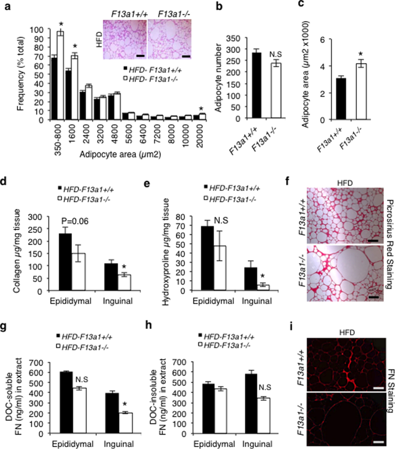
Figure 4. Larger adipocytes and decreased extracellular matrix accumulation in WAT of F13a1−/− mice fed HFD.
(a) Frequency distribution of adipocytes in F13a1−/− and F13a1+/+ mice on HFD diet; the frequency of small and larger adipocytes were increased in F13a1−/− mice. Inset-Hematoxylin and Eosin (H&E) stained sections of epididymal fat pads show larger adipocytes. (b) The total adipocyte number was not increased. (c) An average adipocyte area with image of largest adipocytes found in histology shows significant increase in size. 5–6 fields per mice were counted. (d,e) Sircol and hydroxyproline assay for the collagen content of total protein extracts of epididymal and inguinal fat pads shows a significant decrease in inguinal fat pads. (f) Picrosirus red staining for collagen in epididymal fat pad. (g) (Deoxycholic acid) DOC-soluble and DOC-insoluble FN levels in epididymal and inguinal fat pads show a decrease in inguinal depot. (h) FN levels in DOC-insoluble fraction. (i) Immunofluorescence staining of FN in epididymal fat pad. All error bars represent SEM; Scale bar equals 1000 μm; (n = 4 mice/group); *p < 0.05; N.S.-Not Significant.

