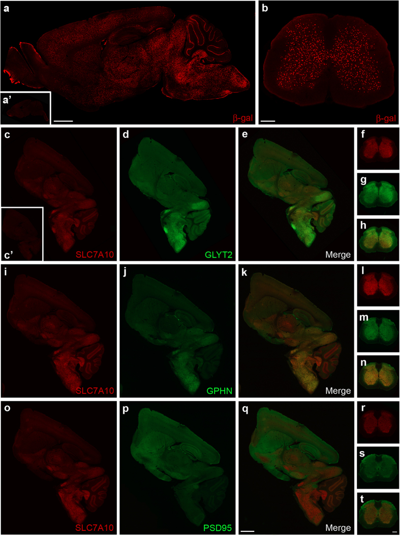
Figure 1. SLC7A10 expression is enriched in the caudal brain, brainstem, and spinal cord.
Using beta-galactosidase (β-gal) as a surrogate marker for SLC7A10 expression, high-density expression is observed in caudal regions of the brain (a) and throughout the spinal cord grey matter (b), as well as in a select population of cells within the cerebellum (a). No beta-galactosidase expression is observed in knockout littermates lacking the targeted Slc7a10 allele (i.e., wild type; a’). Regional distribution of endogenous SLC7A10 expression resembles areas of high density glycinergic inhibitory activity. Endogenous SLC7A10 distribution is similar to that of the presynaptic glycinergic marker GLYT2 in the brain (c–e) and spinal cord (f–h) and similar to that of the postsynaptic inhibitory marker gephyrin (GPHN) in both brain (i–k) and spinal cord (l–n). SLC7A10 expression appears complementary to post-synaptic density protein 95 (PSD95), a marker of excitatory glutamatergic synapses in brain (o–q) and spinal cord (r–t). Slc7a10−/− brain shows complete absence of SLC7A10 immunostaining (c’). Scale bars, 1 mm (brain) and 500 μm (spinal cord).

