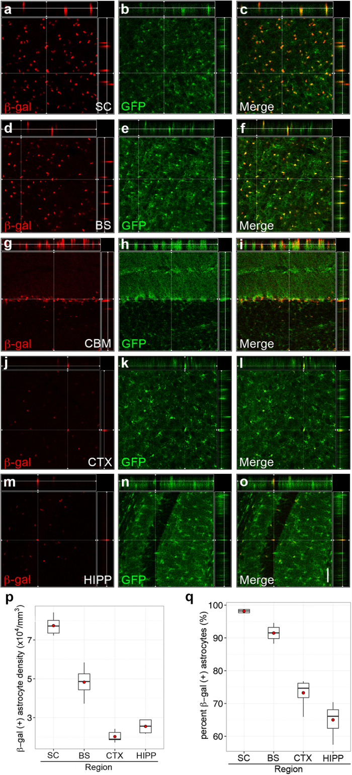
Figure 3. SLC7A10 is enriched in astrocytes in regions of high-density inhibitory activity.

Mice hemizygous for GLT1-GFP and heterozygous for Slc7a10 show beta-galactosidase expression exclusively in GFP-positive astrocytes in the spinal cord (a–c, SC), brainstem, (d–f, BS), cerebellum (g–i, CBM), cortex (j–l, CTX), and hippocampus (m–o, HIPP). Significantly higher densities of beta-galactosidase-positive astrocytes are observed in the spinal cord compared to all other brain regions examined (p < 0.001), and significantly higher densities are present in the brainstem compared to cortex and hippocampus (p < 0.001) (p). Similarly, the percentage of beta-galactosidase-expressing astrocytes is significantly higher in spinal cord and brainstem compared to cortex and hippocampus (p < 0.001) (q). Orthogonal views are shown for each region, demonstrating colocalization of GFP and beta-galactosidase signals. n = 3 biological replicates for each genotype. Scale bar, 50 μm.
