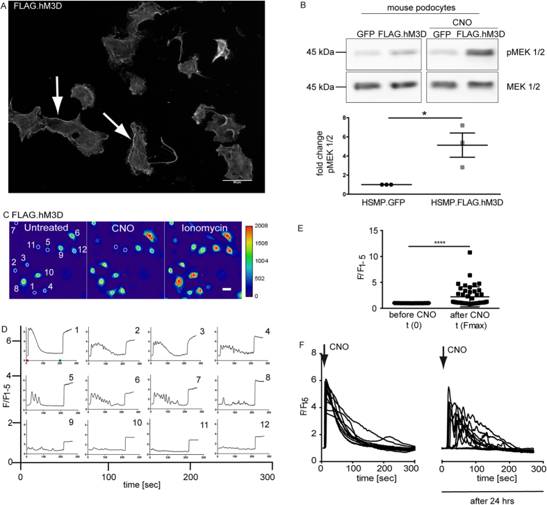
Figure 1. Conditional expression of a functional hM3D in murine immortalized podocytes.
(A) Murine immortalized podocytes stably expressing the FLAG.hM3D were stained with a FLAG antibody to confirm expression and membrane localisation (scale bar = 50 μm). (B) Immortalized mouse podocytes stably expressing FLAG.hM3D or GFP were treated with 1 μM CNO for 10 min. Western Blot analysis revealed a significant increase of the phosphorylation of the MAP-Kinase MEK1/2 after CNO treatment in the receptor expressing cells. (t-test: *p < 0.05). (C,D) Representative images of Ca2+ imaging with Fluo-8 (scale bar = 20 μm). FLAG hM3D expressing podocytes were used for Ca2+ imaging with Fluo-8. Administration of 2 μM CNO leads to an immediate increase of intracellular Ca2+ levels. The time point of CNO administration is indicated with a red tick in panel one. Ionomycin was used as positive control (indicated with a green tick in panel one). 12 cells are depicted from which some reached saturation of the fluorescence signal. Fluorescence is depicted as F/F1–5, where the mean value of the first five measurements (F1–5) prior to CNO stimulation was used for normalisation. Experiments were performed in three biological replicates. (E) Statistical analysis revealed a significant increase of intracellular Ca2+ after CNO stimulation. 45 cells from three biological replicates were included in the statistics. (t-test: p < 0.0001). (F) FLAG.hM3D expressing podocytes were used for repetitive Ca2+ imaging with Fluo-8. Podocytes were treated with CNO at 0 and 24 hrs. At both time points an immediate increase of intracellular Ca2+ levels could be observed. Fluorescence is depicted as F/F1–5, where the mean value of the first five measurements prior to CNO stimulation was used for normalisation.

