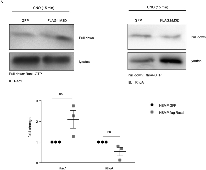
Figure 2. Increased intracellular Ca2+-levels after CNO treatment cause Rac1 activation.
(A) GTPase pull-down assays were performed for active RhoA and Rac1. FLAG.hM3D expressing podocytes and GFP expressing control podocytes were stimulated with 1 μM CNO for 15 min. Pull down of active Rac1 and RhoA revealed no significant changes of active Rac1 or RhoA in FLAG.hM3D-expressing cells in comparison to control cells. Active Rac1 and RhoA levels were normalized to tubulin. (two-tailed t-test: p > 0.05).

