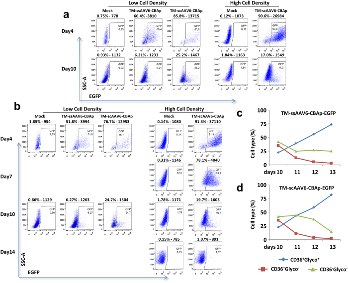
Figure 3. Transduction efficiency of TM-ssAAV6 and TM-scAAV6 vectors in primary human CD34+ cells.
(a) Primary human cord blood-derived CD34+ cells were either mock-transduced, or transduced at day 0 at low (0.5 × 10⁶ cells/ml,) or high (1 × 10⁷ cells/ml) cell density with 20,000 vgs/cell of the indicated AAV6 vectors in serum free XVIVO20 medium. Two hrs later, cells were diluted to 5 × 10⁵ cells/mL and switched to the expansion medium (IMDM + FBS + SCF + IL3 + Epo+ Dexamethasone + β-estradiol + β-mercapthoethanol). EGFP expression was determined by flow cytometry at day 4 and day 10 post-transduction. (b) Following mock-transduction, or transduction of CD34+ cells as described above, cells were switched to the expansion medium for 10 days, and cultured in an erythroid differentiation medium (IMDM + BSA + Insulin + Transferrin + Epo) for an additional four days. EGFP expression was determined by flow cytometry. (c,d) Vector-transduced CD34+ cells cultured in the differentiation medium were stained with hCD36-PE and hGlycophorin A−FITC and analyzed by flow cytometry for the following: non-erythroid (CD36−/glycoA−), and erythroid cells (CD36+/GlycoA+) from day 10 to day 14.

